4.4.5 Classical Swine Fever (CSF), Hog Cholera
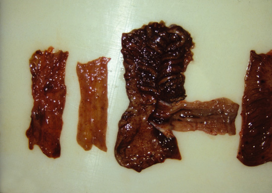
In CSF, haemorrhagic to necrotizing enteritis can be one of the clinical signs in sub acute to chronic cases. This pestivirus infection is highly contagious and is one of the reportable diseases from OIE list A. This economically important disease cannot be ignored when listing the differential diagnosis. Lesions in ileum and colon are indistinguishable from those which occur along with the necroproliferative/haemorrhagic enteritis (see Pict. 4.4.5 a).

Picture 4.4.5 a (by J. Pohlenz)
Haemorrhagic to fibrinous Enteritis in small and large intestine from a fattening pig experimentally infected with hog cholera virus: small intestine at the right side, parts of ileum, caecum, and colon in the middle, and 2 colonic samples at the left side.
CSF has not been diagnosed in the United States of America for many years, but there were several costly outbreaks of this disease in Europe recently. It is essential for people involved in pig disease programmes to know about CSF, since this disease can occur unexpectedly in any country.
Diagnostic methods for the detection of Classical Swine Fever (CSF) virus:
Unlike in acute CSF, it is impossible to make a clinical diagnosis in cases of chronic CSF, as clinical signs and pathological lesions are widely variable and not specific for CSF. Furthermore, concurrent infections with other pathogens often impede a diagnosis based on investigating live animals.
For laboratory diagnosis, the detection of viral antigen and genome, the isolation of virus or the detection of antibodies are performed in selected laboratories.
When using samples from clinically diseased pigs, direct virus detection is recommended. For virus isolation, peripheral blood leukocytes or tissue preparations from tonsil, spleen, kidney and ileum can be incubated on swine cell cultures. Virus isolation in combination with immunofluorescence staining is considered as being the gold standard for CSF virus detection. Alternatively, a molecular biological method
(RT-PCR) or an antigen detecting ELISA can be used. Starting with freshly frozen tissue sections, a direct fluorescent antibody test (FA) results in faster preliminary information compared to the virus propagation in cell culture
but its diagnostic safety is limited.
Antibody detection represents a useful tool for the screening of suspected farms. Different methods are available based on ELISA techniques, immunofluorescence inhibiting assays (IFA) and neutralisation immunofluorescence assays (NIA). ELISA CSF positive or suspected sera have to be retested in the IFA or NIA due to the closely antigenic relationship between CSFV and further pestiviruses like BVDV and BDV.
© Boehringer Ingelheim Animal Health GmbH, 2006
All rights reserved. No part of this Technical Manual 3.0 may be reproduced or transmitted in any form or by any means, electronic or photocopy, without permission in writing from Boehringer Ingelheim Animal Health GmbH.


