

4.4.3 Swine Dysentery
A common disease in the age group of 6 to 20 weeks is Swine dysentery. The causative agent is Brachyspira hyodysenteria. In rare case Brachyspira pilosicoli may be found. Infections with Brachyspira are restricted to the large intestine and do not affect the ileum or parts of the upper small intestine, an important differentiation of swine dysentery against necro-proliferative Ileitis.
In swine dysentery, pigs develop within a few days of infection mucoid to slightly bloody diarrhoea in which the admixtures of coagulated mucus strands become evident within the first week of infection. Later the condition changes to foully smelling watery diarrhoea, where fibrin and blood coagula are contained in the faeces. Pigs rapidly loose weight, but sudden deaths are rare. The infection is usually acquired from other fattening pigs or older breeding stock, and, in contrast to Lawsonia infections, generally 30 to 50% of a group are sick from watery diarrhoea.
Upon necropsy there is a Typhlocolitis with thickening of the mucosal folds present. An intense inflammatory response results in reddening of the mucosa, which is covered by intensely secreted mucus and some fibrin (see pict. 4.4.3a). In later stages a severe ulcerative to necrotizing colitis is characteristic (see pict. 4.4.3b).
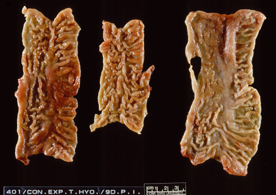
Picture 4.4.3 a (by J. Pohlenz)
Three examples of colonic mucosa with lesions due to Brachyspira hyodysenteriae, 9 days post experimental infection.
Moderate fibrinous colitis with thickened intestinal folds and mild focal ulceration.
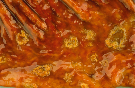
Picture 4.4.3 b (by J. Pohlenz)
Severe fibrinous to ulcerative Colitis, 20 days post infection with Brachyspira hyodysenteriae.
Histological lesions have some similarities with Lawsonia intracellularis infections. In the early phase (6 to 10 days) there is a moderate to severe crypt proliferation in deeper crypts.
The crypt architecture is less intensely altered, crypts are dilated and from the mid crypt region towards the lumen goblet cells are still contained in the intestinal mucosa. Some surface cells are sloughed into the lumen (see pict. 4.4.3 c). In more severe cases there is necrosis on the surface and more intense inflammatory response in the intestinal wall (see pict. 4.4.3 d).
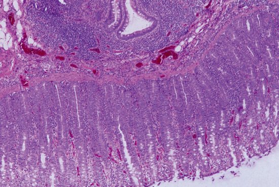
Picture 4.4.3 c (by J. Pohlenz):
Histological section of colon; crypt proliferation in base of crypts, crypts are elongated with immature enterocytes; goblet cells are shifted luminal to mid crypt region. There is a mild inflammatory response in the lamina propria and a lymphatic hyperplasia of sub mucosal lymphoid glandular complexes, 9 days p. i., H&E stain, (~125x).
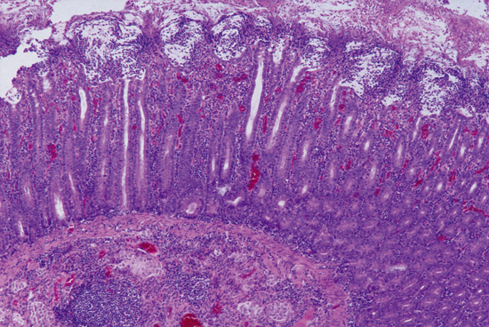
Picture 4.4.3 d (by J. Pohlenz)
Histological section of proximal colon nine days p.i. with Brachyspira hyodysenteria: Crypt proliferation and dilatation of crypts, filled with debris. Severe necrosis on surface; moderate cellular infiltration in lamina propria and sub mucosa.
To detect the organism in histological sections, the silver stain (Warthin/Starry) has been efficiently used for many years (see pict. 4.4.3 e). Cork screw like black stained bacteria are present in the lumen of dilated crypts. Only occasionally Brachyspira spp. are seen in dilated goblet cells, in contrast to Lawsonia intracellularis, which is intracellular in proliferating immature enterocytes. In tissues of pigs infected with Brachyspira pilosicoli, the lesions are less intense, but silver staining is positive.
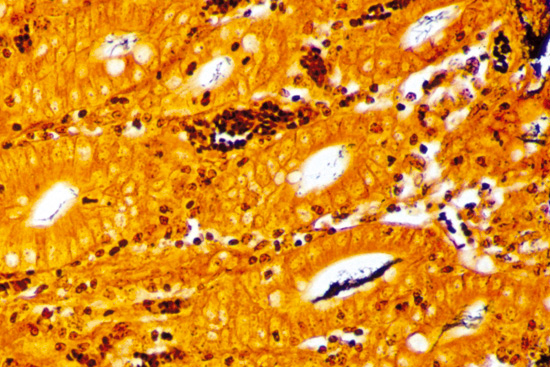
Picture 4.4.3 e (by J. Pohlenz)
Silver staining (W&S) of a colonic section of a pig, infected with Brachyspira hyodysenteria: Cork screw-like black stained bacteria in the lumen of crypts (~500x). Note that Lawsonia intracellularis will also be stained by this method. For clear differentiation use IHC with specific monoclonal antibodies.
Diagnostic methods for the detection of Brachyspira spp.:
In fresh samples from abnormal mucosa or faecal swabs, Brachyspira can directly be examined microscopically under dark ground illumination and phase contrast. In addition, preparations specifically stained are used to demonstrate the presence of Brachyspira. Propagation of Brachyspira can be done requiring cultivation within 24 hours after sampling.
Differential PCR methods can be used to confirm an isolate or to diagnose and genotype Brachyspiraspp. directly from clinical specimens.
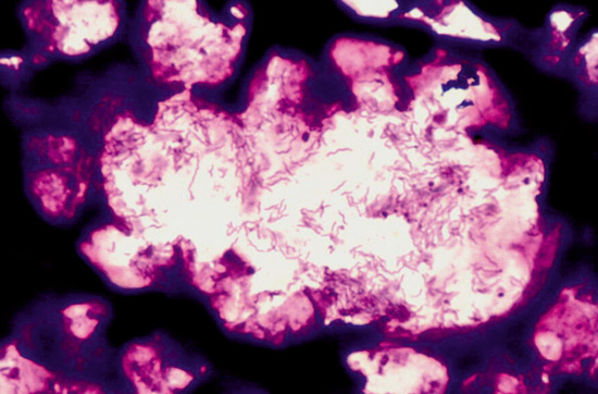
Picture 4.4.3 f (by M. Dünser)
Histological section of a colon crypt with Brachyspira Hyodysenteriae in the lumen of it.
© Boehringer Ingelheim Animal Health GmbH, 2006
All rights reserved. No part of this Technical Manual 3.0 may be reproduced or transmitted in any form or by any means, electronic or photocopy, without permission in writing from Boehringer Ingelheim Animal Health GmbH.