


4.2.1 Chronic Ileitis
The lesions of chronic Ileitis occur most commonly in the terminal 50 cm of the small intestine (see pict. 4.2.1 a) and the upper third of the proximal colon, including the caecum. The magnitude of the proliferation varies widely, but in the advanced lesions the wall is visibly thickened and the overall diameter markedly increased. The affected mucosa is thrown into deep folds, longitudinal or transverse (pict. 4.2.1b). Because of the thickening of mucosal folds in association with proliferation, this condition has been denominated intestinal adenomatosis. The mucosal surface is moist but not mucoid. On the surface, there are flecks of inflammatory exudates, loosely adherent, admixed with fibrin. Peyer’s patches may be indentated. In minor lesions, the area of the terminal ileum, 10 cm proximal to the ileocaecal valve should be examined, as this is the most likely site of a mild infection (pict. 4.2.1c). Care is needed to distinguish minor lesions from contracted mucosa over the Peyer’s patches.
In some pigs with Ileitis, a more severe form can develop where secondary infections such as Fusobacterium necrophorum, Actinomyces spp., or Bacteroides spp., and inflammation occurs on top of the basic chronic proliferative lesion.
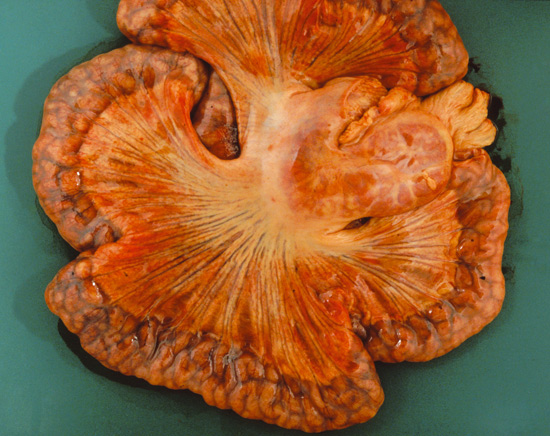
Picture 4.2.1 a (by J. Pohlenz)
Severely thickened ileal wall with congested lymphatics and enlarged mesenteric lymph nodes of a fattening pig with Lawsonia intracellularis infection.
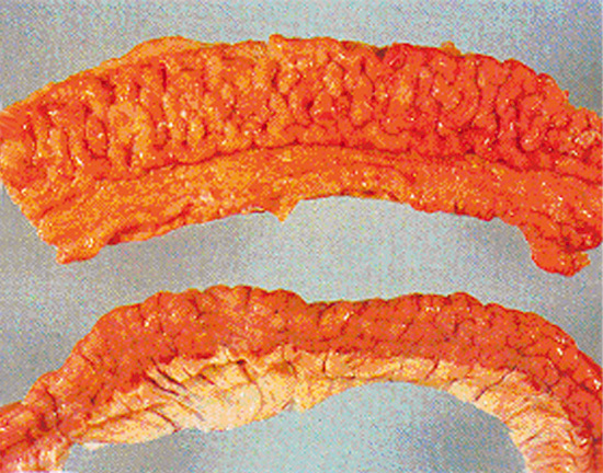
Picture 4.2.1 b (by J. Pohlenz)
Severe serosal oedema (upper part of picture). Marked thickening
of ileal mucosa with bulging of proliferated longitudinal and transverse folds (lower picture of opened piece of ileum).
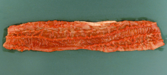
Picture 4.2.1 c (by J. Pohlenz)
Early stage of Lawsonia intracellularis infection in lower ileum. Proliferating longitudinal and transversal folds are protruding into the lumen. The diffusely hyperaemic mucosa is covered by greyish exudates.
This form is called necroproliferative enteritis or necrotizing proliferative enteropathy (NPE) (Picture 4.2.1 d).
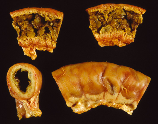
Picture 4.2.1 d (by J. Pohlenz)
Two longitudinal cuts, one crosscut, and one sample of unopened ileum with advanced severe NPE. The lumen is filled with necrotic debris; the intestinal mucosa is thickened and intermingled mucosal cysts.
At autopsy, there is coagulative necrosis with marked inflammatory exudates superimposed on an established lesion of Ileitis. These exudates are a yellow-grey cheesy mass that adheres tightly to the mucosa and may closely follow the original thickened mucosa pattern. Remnants of proliferative epithelium in the deep layers will confirm the diagnosis of Necrotic Enteritis due to Ileitis. In a small number of pigs with Ileitis, the ileal wall becomes firm and fibrotic, but often the mucosa atrophies in these cases. Due to the fibrosis in the intestinal wall, this form has been called Regional Ileitis, or “garden hose gut”, but this condition is become extremely rare.
© Boehringer Ingelheim Animal Health GmbH, 2006
All rights reserved. No part of this Technical Manual 3.0 may be reproduced or transmitted in any form or by any means, electronic or photocopy, without permission in writing from Boehringer Ingelheim Animal Health GmbH.