AHVLA Report: Neonatal Diarrhoea, Osteoporosis and Inclusion Body Rhinitis in UK Pigs
UK - The Animal Health Veterinary Laboratories Agency (AHVLA) has looked into neonatal diarrhoea due to rotaviral and clostridial enteritis, inclusion body rhinitis with and without systemic involvement, multiple diagnoses of disease due to Streptococcus suis and osteoporosis leading to pathological fractures in weaned first-litter sows, according to its Pig Disease Surveillance Monthly Report for April 2015. 3 August 2015
 18 minute read
 By:
By: Increase in salmonellosis due to monophasic Salmonella Typhimurium-like variants
There was an increase in the GB rate of diagnosis of salmonellosis in this quarter from 11.2% in the first three months of 2014 to 17.8% in the same period in 2015 as shown in Figure 1. This was due to an increased rate of incidents due to monophasic Salmonella Typhimurium-like variants (4,12:i:- and 4,5,12:i:-).The diagnostic rate of incidents due to Salmonella Typhimurium (STM) remained fairly constant and of incidents due to other (non-STM, non-monophasic) Salmonella was reduced compared to Q1, 2014. Figures 1 and 2 illustrate this. This was the first quarter that the monophasic variants were responsible for a greater proportion of disease incidents than S. Typhimurium reflecting the increasing prevalence of these variants in the pig population since 2010.
Figure 1: Annual trend in GB incidents of salmonellosis due to monophasic variants as % of diagnosable submissions
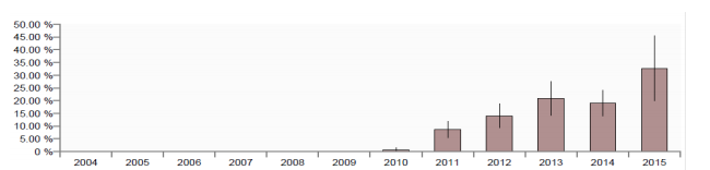
Figure 2: Annual trend in GB incidents of salmonellosis due to STM as % of diagnosable submissions
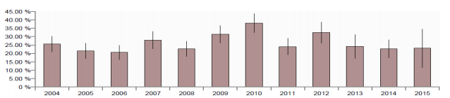
Salmonella 4,12:i:- has typically been less common than the S. 4,5,12:i:- variant. However, reports of S. 4,12:i:- increased almost six-fold compared to the first quarter of 2014 while reports of S. 4,5,12:i:- and S. Typhimurium remained stable, making Salmonella 4,12:i:- the most common serovar isolated from pigs in the first three months of 2015. Phage type U288 was found in 83% of the S. Typhimurium incidents whilst DT193 was found in 71% of the Salmonella 4,12:i:- incidents and all of the Salmonella 4,5,12:i:- incidents.
It is of note that in 60% of salmonellosis incidents diagnosed in pigs between 2005 and 2014 in APHA post-mortem examinations, at least one other disease was also diagnosed indicating that salmonellosis in post-weaned pigs is often part of more complex disease and emphasising the need for comprehensive diagnostic investigations in more severe, unusual or non-responsive disease outbreaks. Some examples of salmonellosis outbreaks are described in this report.
Reproductive disease
No infectious cause established for abortion outbreak
Extensive testing of two aborted litters submitted to the University of Bristol did not detect an infectious cause for a small abortion outbreak in a 460-sow indoor herd. Five sows had aborted or had weak, premature piglets and infertility had been a problem with regular returns. Sows were vaccinated against erysipelas, parvovirus, and porcine reproductive and respiratory syndrome (PRRS). No gross pathology was evident though in both litters there was some variation in size of the piglets, and none were mummified. Testing for PRRSv, pathogenic Leptospira, swine influenza and bacteriology did not provide a diagnosis were unrewarding and foetal histolopathology only showed some evidence for a bacterial inflammatory process in the lungs.
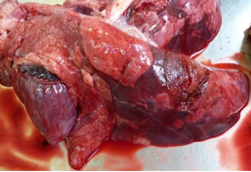
A seven-week-old pig submitted at the same time from this farm had a severe bronchointerstitial
pneumonia identified histologically, consistent with bacterial pneumonia secondary to a viral challenge (Figure 3).
Figure 3: Patchy ventral pulmonary consolidation in bronchointerstitial pneumonia
Tests for swine influenza and PRRS were negative in this pig, however serology on aborting sows showed positive titres which were highest to H1N2, to which one sow had a titre of 1280 and another 2560. This does not confirm involvement of swine influenza as there were no paired sera to demonstrate a temporal association of seroconversion and the reproductive disease. However, previous outbreaks of acute swine influenza in pregnant sows have occasionally also caused small outbreaks of late stage abortion and stillbirths due to the maternal effect of the viral infection and two sows were also seen to be dyspnoeic in the dry sow yards at the time of the abortions. It was agreed that if respiratory disease continued, that further cases would be submitted.
Enteric Disease
Rotavirus causing diarrhoea prior to weaning in small herd and in neonates in commercial herd
Rotavirus was identified as the cause of the diarrhoea in faecal samples submitted to Starcross to investigate diarrhoea in six-week-old preweaned piglets in a Saddleback herd. Porcine epidemic diarrhoea virus (PEDv) was not detected in routine testing for surveillance. PED outbreaks have now been reported in several EU countries but not the UK, the outbreaks in the EU have involved a reportedly milder PEDv strain (INDEL, OH851-like) which differs from those known as virulent from North America and Asia. APHA are testing samples submitted from pigs with diarrhoea for PEDv using PCR and BPEX are funding this testing for units in England. High mortality in sucking piglets with diarrhoea would be of particular concern and should be promptly tested for PEDv. While testing takes place, strict biosecurity measures should be implemented to help limit spread of the virus and there is advice available on the BPEX website http://development.bpex.org.uk/R-and-D/Pig-Health/pedv.aspx.
Neonatal diarrhoea affecting about 50% of recent litters was investigated in a herd which had diagnosed rotaviral enteritis a year previously. The rotavirus infection had been managed by controlled exposure in late pregnancy which had worked fairly well until recently. The diarrhoea was described as watery, starting from two-days-old and affected sow and gilt litters. Three acutely affected live piglets were submitted to Thirsk for investigation. The findings were consistent with neonatal enteritis with watery yellow floccular contents in the large intestine. Rotavirus was detected using PAGE testing and histopathological findings in the intestines supported the diagnosis.
Clostridial enteritis outbreaks continue in neonatal piglets
Two incidents of clostridial enterotoxaemia were diagnosed at Bury St Edmunds. In the first, live threeday-old piglets with diarrhoea were submitted from an outdoor breeding unit on which 10 of a batch of 150 litters were affected with malodourous yellow watery diarrhoea. Affected litters were from any parity of sow and about half of the piglets in these litters were affected. All three piglets had marked necrosis mainly affecting the jejunum and histopathology confirmed an acute necrotising enteritis. Clostridium perfringens alpha toxin but not beta toxin was detected in small intestinal contents suggesting type A enterotoxaemia, however beta toxin is labile and, given the necrosis, it was thought more likely that this was type C disease. No other enteropathogen was detected, including no porcine epidemic diarrhoea virus, and there was no evidence of hypogammaglobulinaemia in the piglets.
In the second incident, three dead five to seven-day-old piglets were examined to investigate diarrhoea and death from four to five-days-old in gilt litters on an outdoor breeding unit. About 20 of the batch of 200 gilt litters were affected and about 60 piglets had died. Severe acute necrotic enteritis was present in all three piglets and clostridial disease was suspected. This was supported by the detection of Clostridium perfringens alpha toxin in small intestinal contents from two of the piglets. When clostridial disease occurs in slightly older piglets as here, concurrent enteropathogens may be playing a role; none were detected and no coccidial oocysts were detected by histopathology or faecal oocyst counts.
Diagnosis of coccidiosis due to Isospora suis in young piglets can sometimes be problematic as oocyst output may be low; submission of live affected piglets, when possible, allows the intestines to be fixed within minutes of death and the coccidial forms can be detected by histopathology.
Salmonellosis outbreaks, some with concurrent disease
One hundred of 600 11-week-old pigs were affected with diarrhoea and wasting with 30 recent deaths on an indoor-nursery-finisher unit. Necrosis of the colon was seen in on-farm post-mortem examination and a sample of colon was submitted to investigate suspect salmonellosis. A monophasic Salmonella 4,5,12:i:- phage type 193 isolate was isolated, supporting this diagnosis. Incidents of salmonellosis in pigs have increasingly been due to monophasic Salmonella rather than S. Typhimurium.
The same Salmonella type was found to be the cause of diarrhoea in a group of 900 seven-week-old pigs, 150 of which were showing diarrhoea, with some coughing and lameness and 20 deaths. In the batch of pigs submitted, two other pigs had streptococcal disease due to Streptococcus suis type 7. One of these had a fibrinous pericarditis, the other had non-specific lesions suggestive of septicaemia and S. suis type 7 was isolated from the lungs and meninges respectively.
Similar mixed enteric and systemic disease was diagnosed in nine-week-old pigs submitted from an indoor nursery-finisher unit to investigate wasting and diarrhoea in approximately 5% of 1500 pigs.
Citric acid in water had recently been stopped and the pigs were being treated with doxycycline in-water for suspected Glässer’s disease. Both submitted pigs were in poor body condition suggesting disease was not acute. One had a dilated, thickened large intestine with confluent diptheresis in the caecum, becoming multifocal in the colon and typical of salmonellosis which was confirmed by isolation of Salmonella Typhimurium phage type U308. Interestingly, an enteropathogenic E. coli (strain E4) was also isolated from the intestine - this may have been involved in earlier enteric colibacillosis. The other pig had a fibrous polyserositis and, in spite of the chronic nature of the lesions, Haemophilus parasuis was isolated confirming chronic Glässer’s disease.
Intestinal obstruction in a smallholder pig
Intussusception of the proximal small intestine associated with long fibre (likely hay) was diagnosed at Shrewsbury in one pig. The pig had died after a short illness and was one of a recently weaned litter of seven pigs on a small holding. There was no evidence of underlying disease and it is possible that this was associated with excessive ingestion of hay at a time when the piglet was undergoing a change in diet after weaning.
Escherichia coli disease in nursery pigs
A nursery unit had increased mortalities from one to three weeks post-weaning although pigs from the originating breeding unit also go to other rearing units where no problems had been recorded.
Affected pigs appeared to go flaccid and develop swollen eyelids. On-farm post mortem investigations did not reveal any clear evidence of internal oedema or oedema of the intestinal tract. One affected pig was submitted to Thirsk and gross findings were largely unremarkable apart from slight oedema of the eyelids and of the subcutaneous tissues of the ventral thorax and abdomen. A profuse pure growth of haemolytic Escherichia coli O139:K82 (E4) was isolated from the small intestine; this isolate is a known cause of oedema disease and enteric colibacillosis in pigs.
Brain histopathology to determine whether lesions consistent with oedema disease were present was not possible as the pig had been shot prior to submission. Interestingly, the group of pigs from which the submitted pig came were being treated with potentiated sulphonamide to which the E. coli isolate showed in vitro resistance.
Respiratory Disease
Actinobacillus pleuropneumoniae involved in respiratory disease outbreaks in finishers
Two outbreaks were diagnosed at Bury St Edmunds. In one, APP was implicated as the cause of increased mortality over the period of a week in 20-week-old housed finishers. Minimal coughing was reported but there were several pigs with dyspnoea; 5% of the group of 800 pigs were affected and 10 had died. Focal consolidation and fibrinous pleurisy was present in one fresh pluck submitted and APP was isolated confirming the diagnosis. No viral involvement was detected and the APP isolated was sensitive to ampicillin. Ampicillin (betalactam) resistance was detected in two of the 12 APP isolates obtained in APHA submissions in 2015 and is a recognised issue in some other pig-producing countries.
In the second, APP was found to be contributing to disease when two 13-week-old pigs found dead were submitted from an outdoor nursery on which 7% of 500 pigs were affected with wasting and diarrhoea and mortality had reached 5%. One pig had a fibrinous polyserositis including polyarthritis and also endocarditis; the other had pleuropneumonia and a fibrous pericarditis. Interestingly, different pathogens were detected in each pig; Haemophilus parasuis was isolated from the pig with polyserositis, Actinobacillus pleuropneumoniae (APP) and Pasteurella multocida were isolated from the other pig, consistent with the lung lesions present. No viral involvement was detected.
PRRS virus sequencing shows likely epidemiological link with another case
Sera were submitted from 12-week-old housed pigs to investigate respiratory disease and high lung pneumonia scores in BPHS (abattoir lesion) reports. The pooled sera were tested by PCR and PRRSv was detected and sequenced (ORF5). The virus detected reflected field challenge and was not similar to live vaccine strains (91.7% and 89.4%) but was very similar (99.8%) to the virus detected in a submission to Bury St Edmunds in February 2015, pointing to an epidemiological link between the two.
Concurrent Glässer’s disease and salmonellosis
Mixed disease was diagnosed in young pigs from a batch-rearing unit with a history of coughing and increased mortality in the preceding two weeks. Post-mortem findings in one pig included fibrin over the abdominal organs, extensive fibrinous pleurisy and a fibrinous pericarditis, suggestive of Glässer’s disease which was confirmed by isolation of Haemophilus parasuis. There was also reddened intestinal mucosa and lymphadenitis in a second pig which had more well-established chronic changes involving the lungs and thoracic cavity. A monophasic Salmonella (4,12:i:-) was isolated from faeces of a third animal which showed similar postmortem findings.
Pleuropneumonia due to Haemophilus parasuis
Haemophilus parasuis was isolated in pure culture from the lungs of a weaned pig. The affected animal was reported to have shown respiratory signs and weakness for a day prior to being euthanased, and was the only affected animal in a group of 350. A pluck was submitted to Shrewsbury for examination and exhibited fibrinous ‘roughening’ of the pleural surface. There was consolidation of much of the lung parenchyma with dark lobules within the dorsal aspects of the caudal lobes and lymph nodes were markedly enlarged.
Inclusion body rhinitis with systemic involvement
Tissues were submitted to Thirsk from three and a five-week-old pigs to investigate the cause of sneezing from three-weeks-old in piglets on a unit with 80 sows. Some were reported to respond to antibiotic treatment while others declined with weight loss and ultimately needed to be euthanased.
Histopathology revealed severe lymphocytic inclusion body rhinitis including lymphohistiocytic portal hepatitis and extensive lymphohistiocytic interstitial/tubulointerstitial nephritis with occasional megalocytic cells with intranuclear inclusion bodies. These findings were consistent with severe cytomegalovirus rhinitis and systemic infection. There was also evidence of secondary bacterial infection and of bacterial colitis in one of the piglets. Previous cases of systemic cytomegalovirus have been diagnosed at Thirsk, with evidence of preceding swine influenza virus infection although that was not identified in this case.
Inclusion body rhinitis as part of a disease complex after weaning
Problems since before Christmas of respiratory disease and wasting affecting around 20% of pigs from two weeks after weaning in a 700-sow herd were investigated by submission of three seven-week-old piglets to Thirsk. Piglets appeared to be in good health and good condition at weaning and were vaccinated against PCV2-associated disease at three-weeks-old. Post-mortem investigation revealed a degree of enteritis in all three pigs, pneumonias of variable severity and upper respiratory tract lesions with the mucosa of the turbinates appearing swollen, reddened and covered with mucus. Further testing, including histopathology, revealed the presence of inclusion body rhinitis and bronchopneumonia involving Actinobacillus pleuropneumoniae and Bordetella bronchiseptica infections. No PRRS or swine influenza viruses were detected. The enteric lesions were found to be due to a combination of rotaviral and E. coli 0157:K,’V17’(V17) infections. The E. coli strain is a recognised porcine pathogen associated with enteric disease. Viral diseases did not appear to be underlying the mixed diseases present and factors affecting disease challenge in the postweaning period including pen hygiene mixing, ventilation, age segregation, and other stresses may be relevant.
Systemic Disease
Multiple diagnoses of disease due to Streptococcus suis type 2
Multiple diagnoses were made, although with differing clinical and pathological findings; several are described here and under nervous disease. In one, six sudden deaths from a group of 700 eight-weekold pigs prompted submission of a dead pig. The pig had a moderate cranioventral pneumonia and Streptococcus suis type 2 was isolated. Streptococcal disease was the cause of death and no viral involvement was detected but lung histopathology suggested earlier swine influenza infection.
When pandemic H1N1 2009 swine influenza emerged in 2009, it was not uncommon for streptococcal disease to occur concurrently with swine influenza.
Another diagnosis of disease due to S. suis type 2 was made when a fresh pluck was submitted to investigate respiratory signs and sudden deaths with poor response to antibiotic treatment in 14-weekold housed pigs. Ten percent of 1200 pigs were reported to be showing respiratory disease, with 15 deaths. There was a vegetative endocarditis affecting the left atrioventricular valve from which S. suis type 2 was isolated. Whilst this pig may have been representative of those dying, it may not have been typical of the respiratory disease and no other respiratory pathogen was identified.
Nervous Disease
Streptococcal meningitis remains a prominent diagnosis in growers
Streptococcus suis type 2 was diagnosed as the cause of cases of meningitis in one pen of 13-week-old rearing gilts, despite S. suis vaccination. Twenty five out of 40 in the pen were affected, with seven deaths. Those treated promptly with penicillin responded well. One gilt was submitted in good body condition and with excess cloudy fluid in both stifle joints: S. suis type 2 was isolated from liver, meninges and joints. On two further farms, S. suis type 2 was also isolated from meningeal swabs collected during on-farm post-mortem examinations performed to investigate sudden deaths and signs of meningitis in six-week-old and nine-week-old pigs.
Two six-week-old pigs were submitted to Thirsk to investigate the cause of lameness and shaking, with some pigs showing slight swelling above the eyes, but not of the eyelids as in oedema disease.
Joints were not obviously swollen and pigs were responding to antimicrobial treatment. Post-mortem investigation revealed evidence of severe polyarthritis, pneumonia, possible septicaemia and meningitis.
There was slight subcutaneous oedema along the ventral body in one pig. S. suis type 2 was isolated from both pigs, but to confuse matters a pure growth of E. coli 0139:K82 (E4) was also isolated from the small intestine of one of the pigs suggesting the possibility of oedema disease.
Although it was suspected that streptococcal infection was the main cause of the problem, as a precaution, brain histopathology was undertaken and confirmed severe purulent meningitis consistent with streptococcal meningitis with no histological evidence of CNS lesions of oedema disease.
In contrast to the above cases involving S. suis type 2, when four-week-old pigs were submitted to Bury St Edmunds to investigate an increase in mortality to between 10 and 15% in the previous three batches of weaned pigs, it was S. suis type 1 which was found to be responsible. The pigs were showing tremors, incoordination and recumbency prior to death. Submitted piglets were in good body condition and had excess turbid yellow fluid in all joints including the atlanto-occipital joint in one pig. S. suis type 1 was isolated from the meninges of these pigs in pure growth and is likely to have been causing polyarthritis as well as meningitis although it was not isolated from the joints.
Musculoskeletal Disease
Kyphosis leading to hindlimb paresis
A 13-week-old pig with hindlimb paresis was euthanased and submitted. There was kyphosis with compression of the spinal column and deformity of the distal thoracic vertebra as shown in Figure 4.
The pig was not dehydrated, had eaten recently and had no abrasions on the legs to indicate long-term recumbency, supporting the history of recent development of the paresis. Pigs such as this with kyphosis (humpy-back) which develop paresis should be promptly culled as recovery is unlikely.
The pig was in a group of 960 in which about 5% were showing lameness with swollen joints and it was recommended that typically affected pigs were submitted to investigate that problem which was not likely to be related.
Figure 4: Spinal cord compression due to severe kyphosis

Osteoporosis leading to pathological fractures in weaned first-litter sows
A number of cases of suspected pathological fractures in first-litter sows occurred shortly after being weaned. A similar problem had occurred a few years ago after which the diet was changed (additional calcium included) and the problem resolved and it transpired that additional calcium was no longer being provided to gilts prior to being served. A typical affected sow which had delivered 16 live piglets and had weaned 10 good piglets was euthanased and submitted to Thirsk having been found off her legs four days after weaning. Post-mortem examination revealed multiple fractures of both humeri and both femurs including the femoral necks and the cortices of the long bones were thinner than expected. It was suspected that the fractures are mainly pathological with the possibility of some degree of trauma contributing after weaning when sows were mixed, particularly as all four limbs were involved. Bone analysis showed normal Ca:P ratios and bone ash which is consistent with lactational osteoporosis as bone is normal in structure but reduced in mass and thus more prone to fracture. The heavy demands on gilts for calcium for both growth and lactation, especially when they have good litters means that osteoporosis is most commonly seen in late lactation or in weaned first-litter sows as on this unit. A shortage of dietary calcium and poor bone reserves predispose to its occurrence and these findings prompted an urgent review of the diet and management of gilts.
Urinary Disease
Chronic kidney disease in infertile gilt in poor body condition
A gilt was euthanased to investigate acute coughing and malaise in a group of 50 10-month-old gilts on an outdoor breeding unit. The gilt was in quite poor body condition and was not pregnant.
There was widespread pulmonary abscessation with fibrous pleurisy and Trueperella pyogenes was isolated. The kidneys were pale and firm to cut and histopathology revealed severe chronic fibrosing glomerulonephritis, most likely due to bacterial infection. Urea levels in aqueous humour correlate well with those in serum and a high urea level was found (30 mmol/l, serum reference range 2.6-8.3) supporting the likelihood of renal failure. The renal and lung disease in this gilt was chronic and she was selected for euthanasia because she was not pregnant and was in poorer body condition that the rest of the group. The reasons for her illthrift were found but not the cause of the acute respiratory disease in the group as she was not representative of the group problem.