

AHVLA Reports Rare Piglet Deformities in June 2014
UK - The Animal Health Veterinary Laboratories Agency (AHVLA) has found a rare type of deformity in piglets, enteric colibacillosis outbreaks in older post-weaned pigs, active swine influenza and porcine cytomegalovirus infections in its June 2014 Surveillance report.Reproductive Disease
Rare type of deformities in piglets due to an undetermined insult early in pregnancy
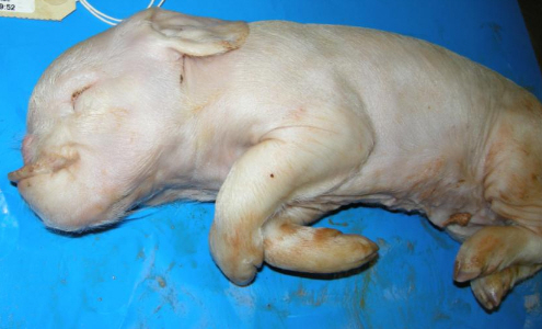
A few piglets with unusual craniofacial deformities were born at term in litters from any parity of sow on an indoor herd in which other piglets in the litters were unaffected. Arhinia was present accompanied by absence of the nasal cavity and olfactory lobes (Figure 1).

The defects were either bilateral or unilateral; in humans this is a rare form of deformity. Based on the timing of embryonic development and the fact that olfactory pits are present in 7mm pig embryos (about 20 days), the histopathologist estimated that any insult leading to arhinia would have been before 20 days gestation.
Thus there was a tight window for causing these lesions and, as disease only occurred over a six-week period, it was transient, although not a point exposure as sequential weekly farrowing batches had a few affected piglets.
Piglets were delivered full-term at the end of May so exposure to the insult would have been early February. The sows were liquid-fed on a wide variety of food by-products from multiple sources which are added to a wheat, barley and soya mix for feeding.
There was no evidence of infectious exposure; and an association between this type of craniofacial malformation and viral infection was considered unlikely. It is possible that the malformations were a consequence of complex interaction between maternal and environmental factors.
Involvement of toxic plants was very unlikely as the sows were indoors. The submission of typical cases in incidents of this nature is very useful in characterising the deformities, exploring possible aetiologies and allowing assessment of the likely timing of any insult.
Alimentary Disease
Enteric colibacillosis outbreaks in older post-weaned pigs than usual
AHVLA have diagnosed a few outbreaks of enteric colibacillosis over recent months in slightly older post-weaned pigs.
The pigs have been dehydrated with fluid enteropathies and culture for E. coli is worth two undertaking for diagnosis if the gross lesions are suggestive of this disease even if the pigs are older than those in which enteric colibacillosis is usually expected.
One of these outbreaks was diagnosed at Thirsk when three pigs were submitted to investigate an ongoing diarrhoea problem affecting pigs at about three-months-old. The pigs were from a small unit weaning at six-weeks of age and kept on solid floored accommodation.
Post-mortem examination revealed enteritis and dehydration and pure growths of haemolytic Escherichia coli 0149:K91, K81 ac (strain Abbotstown) were obtained from small intestinal contents indicating a diagnosis of enteric colibacillosis. No other enteric pathogens expected for this age group were identified. The E. coli strain isolated was a known porcine pathogen associated with enteric disease.
Enteric colibacillosis was also considered the cause of acute diarrhoea affecting six of a litter of 11 eight-week-old Gloucester Old Spot piglets weaned three days prior to submission.
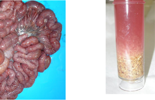
The diarrhoea was brown and watery; one pig died and was submitted to Bury St Edmunds. The pig was markedly dehydrated with a haemorrhagic gastritis and enteritis – the small intestine was reddened and excessively full along its entire length (Figure 2) with watery red-brown liquid and undigested food particles (Figure 3).
A profuse pure growth of an untypeable E. coli was isolated which was sensitive to all antimicrobials in the panel tested. In spite of the E. coli not being typeable, histopathology was typical of enteric colibacillosis and other enteropathogens were not identified. The other affected pigs recovered.

Rotaviral enteritis with possible concurrent clostridial disease in neonatal piglets
Three dead, four-day-old, piglets were submitted for post-mortem examination as part of an investigation into neonatal enteritis. All three pigs had stomachs full of milk, but intestinal contents were watery and pale yellow throughout the small and large intestines.
There was oedema of the spiral colon and the rectum was devoid of content. Rotavirus was detected in the intestinal contents of all three piglets while histopathology revealed acute necrotising bacterial enteritis suggesting a diagnosis of Clostridium perfringens type A although no clostridial toxins were detected and it was considered likely that both these enteropathogens were acting in concert.
Gammaglobulin testing revealed suboptimal colostral transfer in one of the pigs but adequate transfer in the others.
Intestinal torsions in well-grown pigs
An eight-month-old gilt was submitted to Thirsk to investigate ongoing mortality in gilts. The submitted gilt was in good body condition with dark red mucous membranes and a markedly bloated abdomen.
The abdominal cavity contained about 500ml of watery fluid and fibrin strands and both small and large intestines were markedly distended with gas with the small intestine dark-red with port wine coloured contents.
There was a torsion of 180 degrees at the base of the mesentery that included both the small and large intestine. Faeces were of firm consistency. Starcross also diagnosed intestinal torsion as the cause of sudden death of a well-grown pig in a group of 40 growing indoor pigs. Post mortem examination revealed an intestinal torsion around the root of the mesentery.
Excessive build-up of gas in the intestines is a main predisposing factor for intestinal torsion and may occur due to gorging and excessive activity (such as fighting), feeding infrequently or irregularly and other dietary factors such as inclusion of whey in the diet.
Salmonellosis in weaners
Six post-weaned pigs aged between five and six weeks were submitted to Starcross with a history of increased post-weaning mortality. Typical cases developed yellow diarrhoea within three days of weaning and died around five days later.
Twenty-one pigs from a group of 180 had died. Post-mortem examination showed inflammation and necrosis of the gastric mucosa together with thickening and ulceration of the ileal mucosa, which was overlain by a fibrinonecrotic membrane.
A multidrug resistant Salmonella Typhimurium var Copenhagen phage type U288 was isolated by direct culture and advice was provided regarding reducing the risk of zoonotic infection.
Respiratory Diseases
Porcine reproductive and respiratory syndrome underlying bacterial pneumonias
A nursery-finisher unit receiving four-week-old weaners had several batches with unexplained wasting at five weeks of age, which prompted the submission of four pigs. Three of the four had extensive severe pneumonias from which Pasturella multocida and Streptococcus suis were isolated and PRRS virus was detected and considered a significant primary pathogen in the submitted pigs and on the unit.
Active swine influenza and porcine cytomegalovirus infections in fading weaners
Four live piglets, at the point of weaning, were submitted to Thirsk to investigate a problem of fading that started at about six days post-weaning. Between five and seven per cent of piglets were affected with a mortality of about 2½ per cent. Initially at weaning there were no obvious clinical signs but some pigs were reported to then develop respiratory disease and/or diarrhoea.
Piglets were vaccinated before weaning for PCV2, Mycoplasma hyopneumoniae and ileitis. All the submitted piglets had varying degrees of pneumonia and two also had pleuritis. Testing revealed the presence of swine influenza (pandemic H1N1 2009 strain), Streptococcus suis type eight and Mycoplasma hyorhinis and histopathological examination confirmed lung lesions consistent with swine influenza with a secondary bacterial infection.
In addition, there was severe rhinitis which histopathology revealed to be due to Porcine Cytomegalovirus (PCMV) infection and, unusually, there was also evidence of systemic PCMV infection in two of the four piglets.
It is possible that piglets on the unit seen to have diarrhoea were also affected by viral respiratory disease at weaning predisposing to enteric changes as a result of inappetence and possibly dysbiosis and exacerbated by the stresses of weaning.
Severe bacterial pneumonias in late finishers
Actinobacillus pleuropneumoniae and Pasteurella multocida infections were detected in consolidated lungs when 22-week-old finishers were submitted to investigate widespread recurrent respiratory disease.
Coughing and dyspnoea were reported for at least two weeks in housed pigs on a large batch multisource finishing unit with cumulative four per cent mortality since arrival on the unit at around 40kg bodyweight. Various antimicrobial treatments had produced some clinical response but the problem had recurred when medication ceased.
The pigs were vaccinated at weaning for PCV2 and Mycoplasma hyopneumoniae, and for PRRS after arrival at the finishing unit. Despite the appearance of the lungs which was suggestive of viral involvement, none was detected by PCR and immunohistochemistry although the possibility of viral infection weeks earlier in rear could not be ruled out at the time of submission late in rear
Systemic Disease
Thrombocytopenic purpura causing deaths preweaning in several litters
One live and one dead 15- to 17-day-old pre-weaned piglets were submitted to Thirsk to investigate mortality. At the time of the submission four litters had been affected with one to two affected piglets per litter, all within one week. Affected cases were from separate farrowing houses and piglets were either found dead or found lethargic shortly before dying.
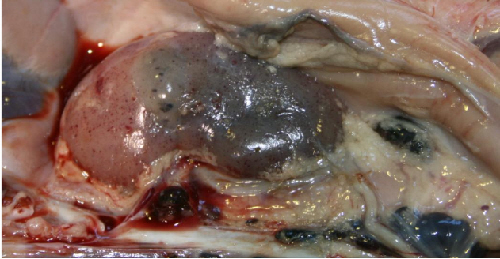
Post-mortem examination revealed widespread serosal ecchymoses and haemorrhages in all the organ systems, including the lymph nodes (Figure 4).
These lesions were not dissimilar to those of swine fever and further information was sought from the attending veterinary practitioner and farm. The lack of pyrexia, absence of disease in gilt litters, fact that sows and other piglets in litter were healthy and specific age occurrence together provided evidence which allowed swine fever not to be suspected. Haematology revealed a very low platelet count (52, reference range 100-900 × 109 per litre).
The overall findings were consistent with a diagnosis of thrombocytopaenic purpura. This occurs when the sow produces antibodies to foetal thrombocyte antigens which are ingested in colostrum.
The antibodies destroy platelets resulting in haemorrhages due to a coagulopathy. The condition only occurs in sow litters following previous exposure of the sow to foetal antigens in the previous litter. A method to control is to avoid breeding sows to the same single boar for subsequent litters although the use of artificial insemination has limited the occurrence of this disease.

Septicaemia due to a less common type of Streptococcus suis
A diagnosis of streptococcal septicaemia was made at Sutton Bonington following examination of an eight-week-old piglet from an open farm. It was the second death in a group of 16 piglets which represented three litters combined. A necrotising sinusitis, fibrinous pericarditis and meningitis was identified.
Bacteriology isolated Streptococcus suis serotype nine in septicaemic distribution, whilst no PRRS or swine influenza viruses were detected.
Porcine circovirus 2-associated disease in vaccinated pigs
Porcine circovirus 2-associated disease (PCVAD) was diagnosed in eight-week-old pigs sourced from two different breeding herds and vaccinated for PCV2 and Mycoplasma hyopneumoniae on arrival at their indoor rearing site at weaning.
The pigs submitted with PCVAD were in poor body condition, one was jaundiced and the other had a bronchointerstitial pneumonia and enlarged inguinal lymph nodes.
Histopathology revealed multifocal granulomatous lymphadenitis and lung lesions typical of PCVAD in both pigs; viral inclusions were visible in lymph nodes of the jaundiced pig and PCV2 was detected by immunohistochemistry in lymph node and lung of the other pig.
Investigations continue into the apparent failure of PCV2 vaccination to control disease and includes PCV2 genotyping – disease was limited to two of the four sources on farm suggesting a possible difference in immunological or virological status of the breeding herds from which the pigs derived.


