

Methods of pregnancy diagnosis
The list below highlights the different methods for diagnosing pregnancy
- Daily observation of the vulva and the behaviour of the female when a boar is present, particularly at 18-22 days post-service.
- Amplitude depth ultrasound machines.
- Döppler ultrasound machines.
- Vaginal biopsy.
- Serum analysis.
- Ultrasound scanners.
1. Daily observations for oestrus
After the eggs are fertilised in the fallopian tubes, the embryos move around the two horns of the womb to become equally spaced. Survival of these to day 10 or 11 starts the pregnancy signal but a failure at this time results in a return to oestrus on a slightly delayed cycle of 22-26 days.
Implantation of the embryo commences during days 12-14 and a minimum of 5 embryos are required for pregnancy to continue. If the pregnancy fails at this time the return to oestrus is delayed to 23-38 days because pregnancy has already started.
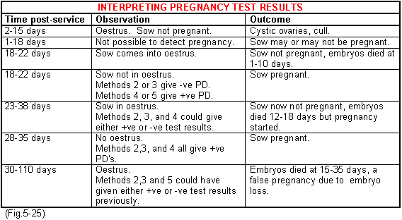
If pregnancy is maintained to the completion of implantation and then fails totally with absorption, the sow becomes pseudo-pregnant for a varying period and then comes through not in pig. In many cases there is a positive pregnancy test reading early on only to find the sow is negative later. The sequence of events and the results you can expect with pregnancy diagnosis are shown in Fig.5-25. Take note that the loss of pregnancy between 15 to 35 days can give false positive test results.

2. Amplitude tests
These machines are only of value from 28-80 days of pregnancy. Beyond this they loose their sensitivity. Also false positives can often be detected if the bladder is full and scanning misses the womb.
3. Döppler tests
Döppler ultra sound machines are more accurate and can be used over the whole range of the pregnancy period from 26 days to term. They are the most popular method with a 90% plus accuracy. The sounds detected in early pregnancy arise from the changes in blood flow that take place in the large arteries supplying blood to the womb. Movement of the foetus and the placenta can also be detected together with the foetal heartbeat. Womb infection, embryo absorption or early oestrus can give false positives and of course wrong interpretations of the sounds or inexperience can give rise to wrong interpretations. Demonstration audio tapes are available with the equipment. The technique is described in chapter 15 Pregnancy diagnosis.
4. Vaginal biopsy
This technique involves the removal of a small piece of the vaginal mucous membrane using a special instrument. The instrument is inserted into the vagina 150-300mm pressed into the membrane and the end manipulated to cut off a small piece. The sample is placed in a small container with a special preservative and posted to a laboratory for histological examinations. It is time consuming, expensive and little used.
5. Serum analysis
This can be carried out after day 22 by using a small stylette to puncture the ear vein. A thin capillary tube collects a spot of blood which is then tested for pregnancy hormones. It is time consuming, expensive and little used. Techniques are being developed to examine faeces to detect pregnancy but as yet are not perfected for commercial use.
6. Ultra sound
Scanning equipment is now available similar to that used in humans for detecting pregnancy. It is expensive but very accurate and can be justified for use on large farms.


