Emerging Threats Quarterly Report – Pig Diseases – July-September 2014
Among the health issues featured in this quarterly report from the Animal Health Veterinary Laboratories Agency (AHVLA) are elevated mortality in Klebsiella septicaemia outbreaks, a changing pattern in the occurrence of PRRS and the continuing threat posed by genotype 2 PRRS virus. 6 January 2015
 17 minute read
 By:
By: Highlights
- New developments in Klebsiella septicaemia outbreaks
- PRRS diagnoses show no summer reduction
- Second systemic porcine cytomegalovirus case in weaners
- Genotype 2 PRRSv remains a threat to GB pigs
Update on Porcine Epidemic Diarrhoea Virus
Following its emergence in the USA in April 2013, the virulent new variant of PEDv continued to cause outbreaks there but with some reduction in cases over the summer months, thought to be, in part, due to the effect of hotter dryer weather conditions on the virus and efficacy of cleaning and disinfection.
The total number of pig farm samples that have tested positive for the Porcine Epidemic Diarrhoea (PED) virus in the USA stood at 8,758 in 31 states at the end of October 2014. The coordinated approach to PED control and stringent biosecurity procedures across the industry are credited with very limited spread of PED in Canada over the summer period although there is concern than outbreaks may reoccur over winter.
The EFSA opinion (EFSA 2014) was published and reviewed what is known of PED viruses present in the EU. Italy and Germany have reported recent outbreaks but these have not had the clinical impact of those seen in North America.
Characterisation of the viruses involved has shown high sequence identity between these and US PEDv strains. Quality Meat Scotland and NFU Scotland are distributing sampling kits to 934 pig units for immediate use if they see clinical signs suspicious of PED. PEDv PCR testing of diagnostic samples submitted to APHA from pigs with diarrhoea has continued through Defra-funded surveillance.
No PEDv PCR-positive samples have been detected in 136 APHA diagnostic submissions from outbreaks of diarrhoea to October 2014. Two outbreaks of diarrhoea in pre-weaned pigs were tested for PEDv by SACCVS and SACCVS has undertaken monitoring testing of more than 900 samples for PEDv/TGEv and deltacoronavirus and none have tested positive for these viruses.
The fact that no endemic PEDv has been identified through this testing supports the view that most of the GB pig population is likely to be susceptible to the virus whatever the infecting strain.
Maintaining surveillance and implementation of the exotic and roundtable recommendations remain key activities for all stakeholders and an another disease awareness item was included in APHA veterinary investigation centre newletters to practitioners.
More information is available on the BPEX web site.
Ongoing Investigations
New developments in Klebsiella pneumoniae septicaemia outbreaks
Consistent with each summer since 2011, Klebsiella pneumoniae subsp. pneumoniae (Kpp) septicaemia outbreaks were diagnosed in July and August. Four outbreaks were diagnosed bringing the total to 14 as shown in Figure 4, involving 12 separate units as two units have had outbreaks recur in a second year.
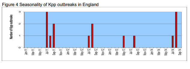
Preliminary analysis suggests that the outbreak Kpp isolates are the same as those involved in outbreaks in previous years; sequence type 25 with a particular combination of a small plasmid and a virulence gene not present in non-outbreak Kpp isolates.
There were three notable developments in 2014. The first outbreak in 2014 was also the first outside East Anglia. An outdoor breeding unit submitted 10 to 12-day-old piglets to APHA Starcross.
Findings were very similar to those seen in previous cases with piglets in good condition showing evidence of recent feeding but with a septicaemic appearance and marked haemorrhages on the intestinal serosa and Kpp was isolated from a wide range of internal sites.
The outbreak was typical of most previous ones, with unexpected deaths of pre-weaned pigs in good body condition. However, mortality was significantly higher at 16 per cent over a period of four weeks. Antimicrobial treatment was instituted at the same time as iron supplementation and, when treated pigs came through, mortality stopped.
This treatment will be continued until the autumn as, so far, Kpp outbreaks have been confined to the summer. The herd has been closed for several years and litters from sows of all parities were affected, the reason for the outbreak is not known and no links to East Anglian units have yet been identified.
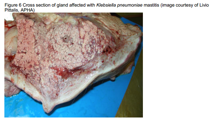
The other three outbreaks were diagnosed in East Anglia during August. These were also typical in causing sudden deaths of piglets from two-weeks-old on outdoor breeding units. However, one outbreak was unusual and significant in that, just after Kpp was diagnosed in piglets on the unit, several sows in the same farrowing batch as the affected piglets became severely ill.
Eight sows were affected from the group of 150 which were soon to be weaned, and five died. Affected sows were acutely depressed and inappetent with skin discolouration especially around the perineum, vulva and mammary glands due to severe mastitis – multiple glands were affected and Kpp was isolated in pure growth. Figures 5 and 6 show the mammary gland lesions.
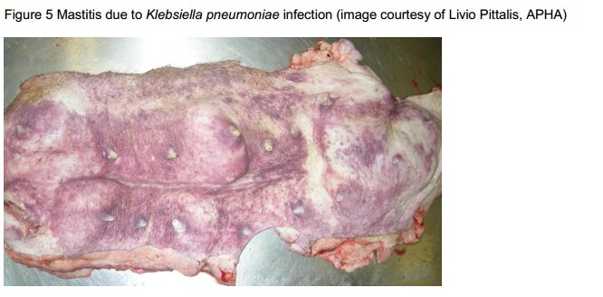
This was the first Kpp outbreak involving disease in sows and prompt treatment of affected sows with potentiated sulphonamide prevented further deaths.
The third development was the first detection of an outbreak Kpp isolate showing acquired antimicrobial resistance. The isolate was resistant to apramycin, spectinomycin, streptomycin, lincomycin-spectinomycin, doxycycline and tetracycline in addition to the innate resistance of all Kpp to ampicillin. The isolate was sensitive to amoxycillin/clavulanic acid, cephalosporins, trimethoprim-sulphamethoxazole, florfenicol and enrofloxacin.
Kpp co-exists in the alimentary tract providing opportunity for acquisition of resistance from other members of the Enterobacteriaceae including Escherichia coli and Salmonella serotypes. Genetic analysis of the resistant isolate and comparison with the sensitive Kpp is to be undertaken to investigate the genetic basis of the antimicrobial resistance.
The genetic analysis of outbreak and historic isolates is also investigating what features of the outbreak Kpp strain may have resulted in its emergence as a new pig pathogen. A presentation was made at the Pig Veterinary Society autumn 2014 conference ('Klebsiella pneumoniae summer outbreaks: What’s new in 2014?' Susanna Williamson and Cornelia Bidewell) to raise awareness and update pig practitioners on the features of Kpp outbreaks and developments seen in 2014.
Unusual Diagnoses or Presentations
There were a number of unusual diagnoses this quarter; details of these have been included in monthly APHA or SACCVS reports. These will be kept under review to assess whether they justify initiation of emerging disease investigations.
Glässers disease with severe skin lesions in weaned pigs
Six-week-old piglets were submitted to Thirsk from a batch of 700 where about 20 had died over a few days. The clinical problem began after weaning with a few pigs being found dead each day. Affected piglets appeared uncomfortable, with swelling of the ventral skin, inguinal area and parts of the limbs.
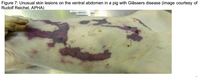
The submitted piglets were not pyrexic but walked with a stiff gait. There was marked pitting oedema of the skin of the ventral abdomen, scrotum and inguinal region and large well demarcated purple areas were present where the skin surface was necrosing as shown in Figure 7. The affected skin was cold to the touch and, on incision, thick subcutaneous oedema was revealed as shown in Figure 4.
There was also evidence of generalised lymphadenopathy and a fibrinous pericarditis, pleuritis and peritonitis, typical of Glässers disease, which was confirmed by isolation of pure growths of Haemophilus parasuis. Subcutaneous oedema and marked scrotal swelling have been reported in past outbreaks of Glässers disease (Hughes, 2003).
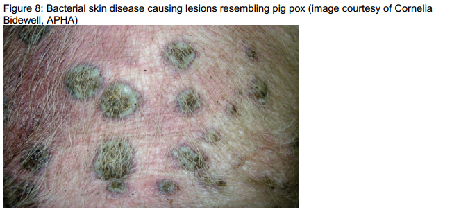
Staphylococcal skin disease causes skin lesions resembling pig pox
Housed finisher pigs were euthanised and submitted as part of an ongoing disease investigation. Clinical signs included pallor, coughing, wasting and 15 per cent mortality. Approximately 200 of 880 pigs were affected over the five-week period and were from a single source. PCVAD had been diagnosed in a previous submission and the virus was a PCV2b typical of previous GB viruses.
The pigs were vaccinated for Mycoplasma hyopneumoniae and should have been PCV2-vaccinated but it was suspected that several batches missed their scheduled PCV2 vaccination. Two of the pigs had multifocal circular raised lesions over the ear pinnae, dorsal neck, body and upper legs measuring 3mm to 3cm with pale peripheries and darker scabbed centres.
Lesions in the more severely affected pig are illustrated in Figure 8. Pig pox was considered a possible differential but pox virus was not detected by electron microscopy and histopathology was consistent with a primary bacterial cause, similar to juvenile impetigo, the pathogenesis of which involves staphylococcal exfoliative toxins.
The pustules were more discrete and the acanthocytes were more numerous than is usual in Staphylococcus hyicus (greasy pig)- associated lesions. Staphylococcus lentus was isolated rather than S. hyicus; this organism is not usually associated with skin disease in pigs and is of doubtful clinical significance.
Changes in Disease Patterns and Risk Factors
Porcine reproductive and respiratory syndrome diagnoses show no summer reduction
Trend analysis of the seasonality of GB porcine respiratory and reproductive syndrome (PRRS) over recent years has shown that there tends to be a peak in diagnoses in the winter months as illustrated in Figure 9 below. This probably reflects the fact that climatic conditions over the winter tend to favour survival of PRRS virus and promote transmission.
Survival of the virus in contaminated pig accommodation, vehicles or on other fomites is also more likely as effective cleaning and disinfection and drying of surfaces is harder to achieve in wet and cold weather. This year, the diagnostic rate of PRRS during July to September was higher than the same quarter in any of the last 10 years.
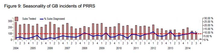
This raises concern that there will be more active infection present going into the cooler months, further increasing the risk of virus spread during this time and contributing to the upward trend in annual PRRS diagnoses illustrated in Figure 10.
Obtaining an accurate diagnosis in respiratory disease outbreaks assists in determining whether PRRS virus is involved and in deciding on specific control measures. Outbreaks of respiratory disease may involve more than one infectious cause and, ideally, a batch of three typically affected pigs early in the course of disease should be sampled or submitted to provide the best material for both diagnosis and pig disease surveillance.
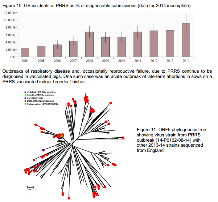
Following mass vaccination of the herd, disease reduced and abortions stopped although increased returns and litters of poor viability persisted, typical of PRRS and causing significant economic impact.
Gilts were home-bred on the unit and their litters were not affected, raising the possibility that endemic virus has been circulating at a low level in the rearing herd, and disease followed spill-over into the breeding herd, affecting mainly older sows. PRRSv sequencing showed the virus to have 89.3 per cent per cent and 87.6 per cent homology to the two licensed vaccine virus strains.
An alert about this trend in PRRS was included in the presentation at the BPEX September producer workshop and the September monthly highlights which are circulated to Pig Veterinary Society, industry and others to increase awareness about the upward trend of PRRS. The alert is found in these highlights via this link [click here].
Porcine circovirus 2-associated disease outbreaks in vaccinated pigs
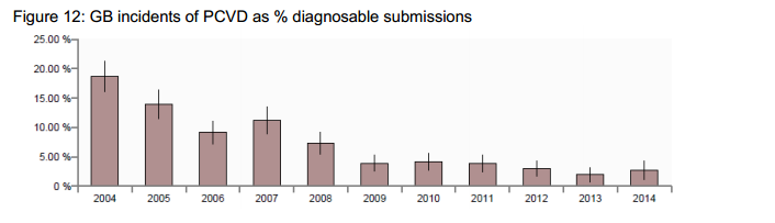
The diagnostic rate of porcine circovirus 2-associated disease (PCVD) outbreaks remains at a low level but there was an increase in diagnoses in Q2 to the highest rate since Q3 2012 and a further increase in this Q3 2014. Figure 12 illustrates the trend in diagnostic rate.
Some of the small numbers of cases seen have been on commercial units vaccinating for PCV2 however, the PCV2 vaccines claims are of a reduction in morbidity of PCVD and scale of disease is important in determining whether vaccine failure may have occurred.
In the most severe outbreak diagnosed, several batches of pigs were thought to missed their PCV2 vaccination and once the problem was diagnosed, the following batches were definitely vaccinated and were not affected. There have also been smaller but significant outbreaks in units where PCV2 vaccination was delayed to between one and four weeks post-weaning.
Interestingly, on multi-source units, disease has sometimes only affected pigs from one breeding source. This may have been a reflection of variability in maternal PCV2 immunity between breeding sources, with variable maternal antibody and, therefore, variable vaccine-take in piglets from those sources.
Joaquim Segalés of Cresa, Spain, gave a webinar on 'Circovirus vaccination and the management of maternally-derived antibodies' on behalf of Zoetis and indicated that high maternal antibody at the time of vaccination was likely to reduce efficacy and, in these herds, later vaccination after weaning may be appropriate.
On units receiving pigs from several sources with piglets of different PCV2 antibody status, the vaccination regime is the same for all sources and thus could provide better protection for pigs from some sources than for others. PCV2 genotyping was undertaken on further 2014 cases and has not detected another PCV2b variant from outbreaks; they have all been typical UK type 2b.
One PCVD outbreak in 2013 involved a PCV2b variant, this has not been detected since. Publication of the findings is planned. No further action is required at this stage but the trend in PCVAD diagnoses will continue to be kept under review.
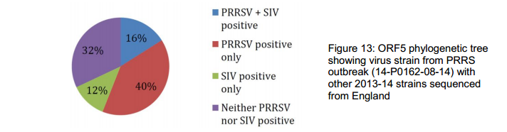
Serosurveillance for PRRS and swine influenza in Scottish slaughter pigs
SACCVS undertook a serological survey for PRRSv and swine influenza virus (SIV) in Scottish pig herds (SACCVS, 2014). Serum samples were collected from ten finishing pigs at slaughter from each of 112 pig herds in Scotland during 2012 to 2013. These herds comprised the majority of units in the Wholesome Pigs Scotland abattoir monitoring scheme/Quality Meat Scotland assurance. Pigs from 16 per cent of herds showed evidence of exposure to both PRRSV and SIV, 40 per cent to PRRSV only, 12 per cent to SIV only and 32 per cent showed no evidence of exposure to either PRRSV or SIV (Figure 13).
The overall seroprevalence to PRRSv of 56 per cent in Scottish pig herds is very close to the seroprevalence of 58 per cent for PRRSv in a UK seroprevalence study on slaughter pigs (Cheney and Powell, 2013). The overall prevalence to SIV of 28 per cent in Scottish pig herds is lower that the 52 per cent herd prevalence reported by Mastin and others (2011), however there were differences in the sampling frame. The Scottish study was conducted in collaboration with Quality Meat Scotland.
Second case of systemic porcine cytomegalovirus infection in weaners
Porcine cytomegalovirus (PCMV) due to a beta herpes virus is occasionally diagnosed in APHA, usually associated with rhinitis, and sometimes wasting, post-weaning. PMCV including systemic lesions was reported in the April to June 'Emerging Threats' report, on that occasion with swine influenza and streptococcal disease.
This second incident of PMCV with systemic lesions was diagnosed in September as the cause of sneezing and ill-thrift in five to 10 per cent of pigs soon after weaning on a breeder-finisher unit. In this case, the PMVC was considered the primary cause of the ill-thrift, although the possibility of earlier involvement of swine influenza is being investigated. Histopathological lesions were present in nasal epithelium (Figure 14) and kidney.
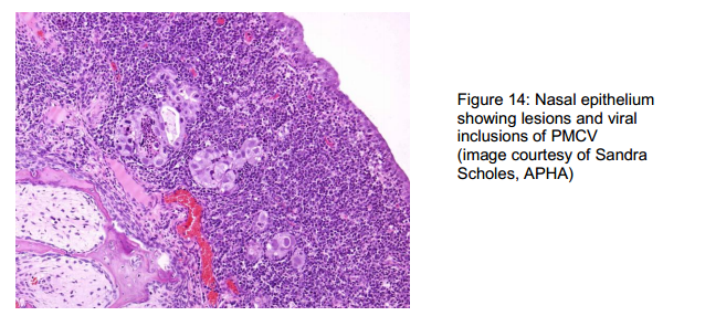
It is likely that PMCV could be underdiagnosed if other more severe diseases are present and it is secondary in clinical significance. Eight PMCV diagnoses were recorded in VIDA over the last 10 years. In three submissions it was the only diagnosis made, in five it was with other diseases (mainly respiratory diagnoses but one with enteric colibacillosis). The primary clinical signs were respiratory or wasting, the secondary clinical signs were respiratory, wasting or diarrhoea.
Pigs were submitted for diagnosis aged between three and five weeks old. Interestingly, a severe outbreak of PMCV in Switzerland with clinical signs of lethargy, respiratory disease, wasting and one stillbirth was described at the joint Europathosurveillance network - European Society of Veterinary Pathologists meeting in Berlin in September 2014 (Marti and others, 2014).
The APHA case was presented at the Pig Veterinary Society autumn conference ('Sneezing pigs'. Duncan Berkshire, Bishopton Veterinary Practice) which will help raise awareness. Exposure of replacement breeding pigs to ropes which have hung in weaner pens is being attempted for control.
Horizon Scanning
African Swine Fever spread in EU in Eastern Europe
During July to September 2014, ASF continued to cause disease and deaths in wild boar in Estonia and in wild boar and backyard pigs in Poland, Latvia and Lithuania and there was continued geographic spread in the region. A large commercial pig unit considered to be biosecure was infected in Lithuania for which the source of infection is not known although an infected wild boar carcase was found nearby.
Biting fly transmission is being assessed by Pirbright as a rare but possible seasonal means of transmission. The herd was culled and there was no spread to other commercial units in Lithuania. ASF in Latvia is of concern as numbers of wild boar and backyard pig cases were reported and some significant geographical jumps have occurred.
Some of the wild boar carcasses were found scavenged and had been dead for a significant time. Each of the infected EU countries have instituted specific control measures to address the outbreak in their wild boar and pigs. It is considered that the risk of ASF would increase significantly if infection spreads into West Poland and/or into commercial pig herds. Up to date preliminary outbreak assessments are available on the UK government web site.
The Pig Veterinary Society held a CPD event which included ASF and CSF to increase awareness amongst veterinarians attending pig units, three speakers from APHA contributed to this day which was well-received.BPEX held a biosecurity workshop for pig producers in September in York to which APHA contributed ('African Swine Fever – what is it?' and 'Surveillance for Threats to GB pigs' Susanna Williamson).
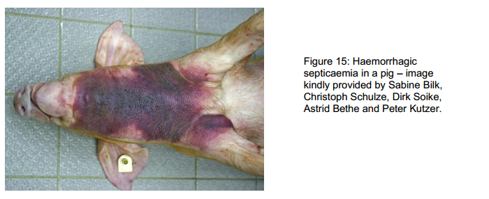
Pasteurella multocida septicaemia as a differential for porcine notifiable disease
Haemorrhagic disease due to Pasteurella multocida septicaemia was diagnosed in pigs in the summer of 2010 for the first time in Germany since 1986 and the outbreaks in pigs since then were described in a poster presented at the joint EPSN-ESVP 2014 meeting (Bilk and others, 2014).
In two herds, mortality was 11 and 14 per cent and the lesions in the pigs are shown in Figures 15 and 16. The haemorrhagic nature of these, distribution and associated subcutaneous oedema and reaction make this disease a differential diagnosis for both swine fevers and anthrax which, in pigs, results in marked throat swelling.
Pasteurella multocida septicaemia outbreaks are occasionally diagnosed in GB pigs but show no increase at present. The results of multilocus sequence typing (MLST) of APHA P. multocida isolates from pigs was reported in the April to June 2014 'Emerging Threats' report and did not detect an emergent strain.
Increased risk of ergot in 2014 cereal crops used for pig feed
Veterinary surgeons attending pigs were asked to be vigilant for signs of ergotism following alerts to cereal growers to monitor their crops following high levels of ergot being found in heavily infested blackgrass areas.
Feed compounders should be aware of the problem and reject cereals containing above a threshold number of ergots in a sample. The presence of ergots does not represent a general mycotoxin problem as a different fungal contamination is responsible.
An alert was sent to pig practitioners and circulated to VIOs raising awareness of this risk and the clinical signs of ergotism. No suspect cases have been diagnosed by, or reported to, APHA or SACCVS.
Genotype 2 PRRSv remains a threat to GB pigs
The threat of North American or genotype 2 PRRSv should not be overlooked while there is high concern about ASF and PED. Genotype 2 PRRSv has never been detected in UK pigs but remains a threat from many other pig producing countries in the world including Europe, North America and Asia.
Immunity to the endemic European or genotype 1 PRRSv would not protect against genotype 2 PRRSv which also tends to be more virulent than genotype 1 virus. Genotype 2 PRRS is specifically mentioned as a threat in the National Pig Association’s voluntary protocol for live pig imports.
Although it is not a statutory requirement, it is vital that pigs imported live are tested prior to import and again while in quarantine to prevent incursion. Pigs can carry PRRSv for weeks to months in their tonsils after recovery from the initial infection and so can appear healthy while still infectious.
The PRRSv PCR used at APHA and SACCVS distinguishes genotype 1 and 2 at the outset which would help early detection if samples are submitted. Investigation at APHA or SACCVS of disease outbreaks in pigs which are severe, unusual or unresponsive disease outbreaks is actively encouraged. The on-line advice provided will also assist practitioners to select suitable samples and tests on the AHVLA web site.
Further Reading
Find out more information about the diseases mentioned by clicking here.
You can view the full report by clicking here.
January 2015