

DISEASE FOCUS - PMWS and PDNS (updated 3rd Feb. 2001)
Mike Muirhead BVM&S, FRCVS, DPM and thePigSite Consultant provides the latest updated information on Postweaning Multisystemic Wasting Syndrome and Porcine Dermatitis and Nephropathy Syndrome.| Postweaning Multisystemic Wasting Syndrome |
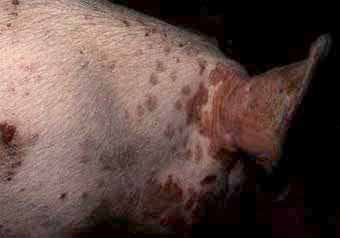
 Skin lesions associated with PMWS and PDNS 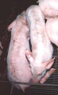 A typical wasting weaner with PMWS 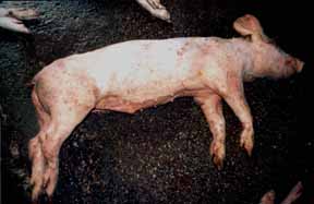 Pig which has died from PMWS
|
PMWS has during the past year or so become of significance and considerable concern in many countries particularly Canada, the US, Europe and the Far East. It is manifest as the name implies by wasting in pigs from around 5-6 weeks of age to around 14 weeks and is now considered a primary disease.
The disease is associated in part with a porcine circovirus (PCV), so called because its DNA is in the form of a ring. It is extremely small and hardy. There are two serotypes, Type 1 causes no known disease. Type 2 can be found in the lesions and can be isolated in pure culture. There are several different strains (biotypes and genotypes). Antibodies to circovirus type 2 have been detected in pig sera collected in Belgium in 1985 but the clinical disease was not described until 1991 in Western Canada. It has since become widespread in North America and Europe.
Young experimentally inoculated colostrum deprived pigs given Type 2 alone sometimes, develop typical lesions. However, they are more likely to develop lesions if another virus, such as porcine parvovirus (PPV) or porcine reproductive and respiratory syndrome virus (PRRSV), is inoculated at the same time. Naturally occurring clinical cases in the field seem always to have a dual infection with PCV Type 2 and some other virus but most pigs which are infected with PCV and PRRS do not develop clinical PMWS.
Serum surveys in Europe and North America have shown that infection has spread widely through the pig population but only a small proportion of seropositve herds have a history of clinical disease. It seems that most infections are sub-clinical. It is not known why some infections result in disease. Piglets may become infected before weaning.
In Western Canada the disease is often reported in high health herds.
Symptoms
Weaners & Growers- PMWS tends to be a slow and progressive disease with a high fatality rate in affected pigs.
- Starting usually at about 6 - 8 weeks of age, weaned pigs lose weight and gradually become emaciated. Their hair becomes rough, their skins become pale and sometimes jaundiced and they are innappetant.
- Sudden death.
- Enlarged peripheral lymph nodes, particularly in between the back legs. Hold the pig up and you may see the inguinal nodes up to the size of golf balls.
- May show diarrhoea in 30% of cases.
- Cases of porcine dermatitis nephropathy syndrome (PDNS) are often seen in herds affected with PMWS.
- May show respiratory distress or laboured breathing caused by interstitial pneumonia.
- Discoloured ears may also be seen.
- Incoordination.
- Nervous signs may occasionally be seen.
- Post weaning mortality is likely to rise to 6 - 10% but is sometimes much higher (20%). In older pigs mortality can rise to 10%.
- Clinical cases may keep occurring in a herd over many months. They usually reach a peak after 6 - 12 months and then gradually decline. Numbers affected vary from one group to another.
- Growth rates in affected pigs are often normal.
- No response to treatment
- Mature animals, sows, boars and sucking piglets are not affected and it is uncommon for newly weaned pigs to be affected before 6 weeks of age.
Causes / Contributing factors
- Infected faeces.
- Mechanical means via clothing, equipment, trucks etc.
- Possibly birds and rodents.
- Circovirus has also been detected in semen from apparently healthy boars.
- It is not known what other ways the virus spreads between pigs or between herds.
- Mixing and stress.
- Continual production.
- High stocking densities.
Diagnosis
 Skin lesions associated with PMWS and PDNS  Enlarged lymph glands associated with PMWS |
- Since most herds have antibodies to PCV, blood testing a herd usually does not help.
- The clinical signs are not specific (although the picture may be highly sugestive) and to make a diagnosis it is often necessary to post mortem several pigs.
- Diagnosis is based upon the presence of PCV type 2 histological lesions in lung, tonsil, spleen, liver and kidney tissues. Immunohistochemistry is used to demonstrate PCV in tissues. Probably many small mild outbreaks go undiagnosed.
- The gross post mortem lesions are variable.
- The carcass is emaciated and may be jaundiced.
- The spleen and many lymph nodes are usually very enlarged, however, the clinical picture with enlarged lymph glands is highly suspicious.
- Kidneys may be swollen with white spots visible from the surface.
- The lungs may be rubbery and mottled with oedema. Microscopically these lesions are characteristic and diagnostic particularly if the circovirus is demonstrated in them. If affected pigs are suspended by their back legs the inguinal lymph nodes appear enlarged often the size of large grapes.
- Oedema or fluid may be seen in the chest and abdominal organs and tissues.
Similar diseases
Many conditions, such as starvation, malnutrition, lack of water, gastric ulcers, enzootic pneumonia, coliform enteritis, swine dysentery, PRRS and other diseases, can cause similar signs. These all have to be eliminated if a specific diagnosis of PMWS is to be made. One disease deserves special mention here, porcine dermatitis and nephrosis syndrome (PDNS) because it can precede, occur at the same time or follow PMWS. The relationship between these two diseases is not known but each can occur in herds without the other being present. (See the latest on PDNS).Treatment
- Antibacterial medication is usually ineffective unless given preventively for a long time in advance of when the start of the disease is anticipated. However, recent reports from Eastern England report good responses to controlling secondary infections using stabilised amoxycillin in feed. (Not a cure).
- There is no vaccine but if a pasteurella is isolated it would be possible to produce an autogenous vaccine.
- Recent reports have indicated that where secondary enteric infections occur the use of tiamulin by both injection and in feed may be useful.
- Pigs are also reported to respond well to injections of corticosteroids (2mg/kg) with improved growth rates and reduced mortalities.
Management Control and Prevention
- Control of the disease is based on pig flow, particularly all-in, all-out systems. Do not move pigs from one batch to another.
- Virkon S has been shown to be effective in killing the virus. PCV is a very persistent virus in the environment.
- Pay attention to good husbandry, ventilation and temperature.
- Avoid high stocking density and reduce mixing of pigs.
- Avoid fostering pigs after 48 hours of age.
- Early recognition of sick pigs and segregation is essential.
- If other diseases are present treatment and control of them may help overall.
- Closure of the herd to build up a herd immunity has been tried but without much success. The suggestion has been made that, since the virus cycles in the nurseries their temporary emptying may break the cycle. It has been shown that groups of young pigs that are removed from the farm and reared elsewhere do much better than those left in the herd.
- Keep similar age groups of pigs segregated to separate buildings or sections and reduce faecal transfers as far as is practicable.
- Use solid partitions between pens.
- It may be worthwhile considering the use of segregated disease. See Chapter 3 - Segregated Disease Control - page 96.
- Vaccinate for parvovirus (PPV) and control PRRS.
- Only purchase breeding stock from herds with no history of the disease or close the herd and use AI only.
- Only use semen from AI studs where all the boar sources have no history of disease.
- Check the biosecurity of the herd, including isolation of incoming stock, entry procedures for people and general hygiene. See Chapter 2 - How Infectious Agents are Spread.
- Pay particular attention to the possibility of faecal transmission particularly by lorries.
Further Information
 |
- The Merial Symposium at the 16th IPVS Melbourne September 2000 reported new management control measures from France including abandoning the following practices:
- Batch mixing.
- Multiple and repeat mixing.
- Litter mixing and distribution of piglets into batches.
- Fostering.
- Open work partitions between pens allowing nose to nose contact.
- Big size pens.
For details on the Merial PMWS Symposium given at the International Pig Veterinary Society Congress, Melbourne 2000 please email: Brian Rice - Merial
- Batch mixing.
- Recent comments by veterinarians in the field1 in the UK suggest that a high proportion of cases of PMWS are associated with corporate production and the mixing of different sources of pigs. This appears to start the spread of the disease by horizonal transmission. Cases are being reported of disease on farms where no pigs have been imported for may years. Birds or mechanical spread have to be considered in such cases.
1 J.D Mackinnon Vet Record Vol 147-5 page 144
- Recent work by Allan and others2 reports the production of disease following inoculations of PCV-2 by the oral/nasal route in colostrum fed pigs that were also vaccinated with Mycoplasma hyopneumonia followed by Actinobacillus pleuropneumonia vaccine. Pigs infected but not vaccinated did not develop disease.
It is postulated by these authors that either the virus has mutated to a more virulent form or changing management factors such as mixing, earlier weaning, multiple sources, vaccinations etc. have contributed to the emergence of the disease.
2 G.M Allan and others Vet Record Volume 147-6 page 170 - The Veterinary Laboratories Agency in the UK has carried out another phone survey of veterinary practices who report 170 new incidents of PDNS. 93% of the cases of PMWS and 95% of cases of PDNS have occurred in the pig dense areas of the eastern region. No cases of PMWS and only one of PDNS have been reported in the East Yorkshire area. This suggests movement of the diseases are related to local spread following their introduction. Only a small amount of pig movement takes place between regions.
The surveys carried out suggest 9% of holdings with 100 sows or more in England and Wales may now be infected.
- Researchers from Iowa have apparently succeeded in reproducing PMWS using only circovirus type 2 (PCV2)
- Abortions, mummified pigs, heart sac infection, PDNS and congenital tremors (trembling piglets) are also thought to be associated with PCV2.
- The virus has also been isolated from semen. The significance of this is yet to be determined. This virus joins the list of others including Parvovirus, PRRS, B.suis, and PRV (Pseudorabies / Aujeszkys disease).
Further reading on PMWS and PDNS
For further information on PMWS and PDNS go to our TECHNICAL ZONE| Porcine Dermatitis and Nephropathy Syndrome |
 |
Veterinary Warning
Dr Stan Done from the Central Veterinary Laboratory in the UK has warned Veterinarians to be on their guard if dealing with the acute epidemic form of PDNS.The symptoms and post-mortem picture are very similar to Classical Swine Fever and include enlarged lymph glands, haemorrhages at any site, cavity, organ or tissue and the presence of fluid in any body cavity.
Such a picture should be immediately reported to the authorities for differential diagnosis
Cause
The cause is unknown. The lesions are in the blood vessel walls throughout and if viewed microscopically suggest a hypersensivity reaction to something in the blood stream, possibly a bacterial toxin. In field studies in Scotland a specific strain of Pasteurella multocida has been isolated consistently from affected pigs whereas the majority of strains isolated from unaffected pigs are different. However, cause and effect have not yet been proven.Some herds in which the disease is occurring have been shown to be infected with porcine circovirus (PCV), which sometimes causes confusion, but PCV is widespread in pig populations and its simultaneous presence may be coincidental. However, porcine post weaning multisystemic syndrome (PMWS) sometimes occurs at the same time as PDNS or precedes it in a herd or follows it. The relationship between these two diseases is not known but each occurs in herds without the other being present. The virus porcine reproductive and respiratory syndrome (PRRS) has been detected in some pigs with PDNS and was suggested as a possible cause because it damages blood vessel walls but, again, cause and effect have not been proved and since PRRS virus is widespread in the many pig populations its presence may also be coincidental. Furthermore, not all herds with PDNS are seropositve for PRRS.
Current thoughts on the cause of PDNS suggest it is an immune complex mediated disease associated with abnormal stimulation of the immune system. This implies antibody antigen reactions. It has been postulated that the condition could be initiated by factors such as medicines, vaccines, chemicals and infectious agents. Recent tests and data have shown that Porcine circovirus Type 2 (PCV2) can be identified in a high percentage of cases the same as PMWS. Further studies are being carried out.
Spread
It is not known how the disease spreads between pigs or between herds or what triggers off a clinical outbreak. However sources of incoming pigs should only be purchased from herds with no history of disease.Clinical signs
- PDNS occurs mainly in growers and finishers, 12 to 14 weeks of age and sporadically in other age groups.
- The most striking sign in live clinically affected pigs is the appearance of extensive purplish red slightly raised blotches of various sizes and shapes over the chest, abdomen, thighs and forelegs.
- Over time the blotches become covered with dark crusts and then fade leaving scars.
- The pigs are depressed and may have a fever.
- They are usually reluctant to move eat, lose weight.
- Sometimes they breath heavily.
- Most pigs with skin lesions die.
- Oedema or fluid may be seen on the limbs and around the eyelids.
- Superficial lymph nodes may be enlarged.
- Diarrhoea in some pigs.
Diagnosis
- The clinical signs are strongly suggestive but not diagnostic. Gross and microscopic post mortem examinations are needed to make a firm diagnosis.
- At gross post mortem examination lymph nodes, particularly those at the rear of the abdomen which are not usually examined, are reddened and enlarged haemorrhagic with fluid.
- There is often fluid in the abdomen.
- The most consistent lesions are in the kidneys which are swollen, pale and mottled with many small haemorrhages showing through the surface.
- Tests can be done for high urea and creatinine levels in the blood which indicates severe kidney damage. These tests may be negative if the kidneys are less severely affected. Microscopically, the lesions in the blood vessel walls are distinctive.
- Since the cause is unknown there are no specific diagnostic tests.
- Gastric ulcers and haemorrhage in the small and large intestines.
Similar diseases
Some outbreaks, clinically and at post mortem examination, resemble Classical swine fever (CSF), (Hog cholera), or African swine fever (ASF), which in most countries are legally notifiable and, if confirmed, herds are slaughtered out. If many pigs with PDNS are subjected to detailed post mortem examination virtually all the typical gross lesions of swine fever and African swine fever are likely to be found, not in a single pig but scattered through them. This causes major problems for the farmer and the vet since the farm and such places as slaughter houses, to which pigs have been delivered, may be closed pending investigation.Fortunately laboratory tests for CSF are rapid and accurate but tests for ASF may take several days to be sure of a negative result.
A special note should be made of PMWS because a number of the clinical signs are very similar and be present at the same time as PMWS. The relationship between these two diseases is not known but each can occur in herds without the other being present. (See the latest on PMWS).
Other diseases which might be confused with PDNS include erysipelas and Actinobacillus suis. Other kidney conditions may also be confused with PDNS.
Treatment
- Antibacterial medication is usually ineffective unless given preventively for a long time in advance of when the start of the disease is anticipated.
- It is a good idea to get a laboratory to isolate bacteria such as Pasteurella form the herd and to run antibacterial tests to provide a more rational choice of preventative medicine.
- There is no vaccine but if a pasteurella is isolated it would be possible to produce an autogenous vaccine.
Management control and prevention
- Isolate incoming pigs for 6 weeks and check the source before entry.
- Control of the disease is based on good husbandry.
- Early recognition and segregation of sick pigs is essential.
- Dissemination of the disease has been associated with the movement of pigs from affected farms.
- Disease has been associated with the mixing of pigs at weaning from multiple sources.
- Lateral movement of disease in dense pig production areas is believed to be a factor.
- Birds are thought to spread desire.
- Fungal toxins and in particular ochratoxin may affect the immune system.
- In finishing units do not buy pigs from infected sources.
- Ideally do not mix pigs less than 30kg weight.
- Keep mixing of pigs and stress to a minimum.
- Adopt all in all out procedures and disinfect buildings between batches.
Further reading on PDNS
 |
For details on the Merial PMWS Symposium given at the International Pig Veterinary Society Congress, Melbourne 2000 please email: Brian Rice - Merial
Also go to our PMWS and PDNS TECHNICAL ZONE


